Introduction to Ovarian Cancer and Early Detection Challenges
Overview of Ovarian Cancer
Ovarian cancer, a formidable adversary in women’s health, stands as the most lethal gynecological malignancy and ranks as the fifth leading cause of cancer-related deaths among females in the United States. Projections for 2022 estimate nearly 19,880 new cases and 12,810 fatalities. The insidious nature of ovarian cancer lies in its silent progression, often eluding detection until it reaches advanced stages. However, if detected early, the 5-year survival rate could soar from a grim 31% to 45% to an encouraging 93% to 98%. The genesis of high-grade serous ovarian cancer, the most common subtype, is now believed to originate within the fallopian tubes (FT), well before the emergence of symptoms, underscoring the critical need for early detection.
Current Challenges in Early Detection
Despite advances in medical science, early detection of ovarian cancer remains a formidable challenge. No universally accepted screening method exists, leaving high-risk women with limited options such as annual transvaginal ultrasound and serum CA125 measurements. Even these methods fall short, with only 50% to 60% of cases detected early in high-risk groups. For women at average risk, the situation is even more precarious, as these screenings are not recommended. The quest for early detection is further complicated by the need for high-resolution, sensitive imaging techniques that can discern subtle pathological changes within the fallopian tubes.
Importance of Early-Stage Diagnosis
The importance of early-stage diagnosis cannot be overstated. Early detection not only dramatically improves survival rates but also opens the door to less invasive treatment options. It allows for timely interventions that can prevent the progression of the disease, significantly enhancing the quality of life for patients. Recognizing the origins of ovarian cancer within the fallopian tubes has shifted the focus of early detection efforts, with the potential to transform the landscape of women’s health care.
Historical Limitations of Diagnostic Tools
Historically, the tools available for the diagnosis of ovarian cancer have been limited by their invasiveness, lack of specificity, and insufficient resolution. Traditional imaging methods, such as ultrasound and CT scans, often fail to detect early-stage tumors. Moreover, the reliance on biomarkers like CA125 has proven inadequate due to low sensitivity and specificity. These limitations have necessitated the development of new technologies capable of providing detailed, real-time imaging of the fallopian tubes, where the earliest signs of ovarian cancer may be found.
Development of the Falloposcope
The Concept and Research Behind the Falloposcope
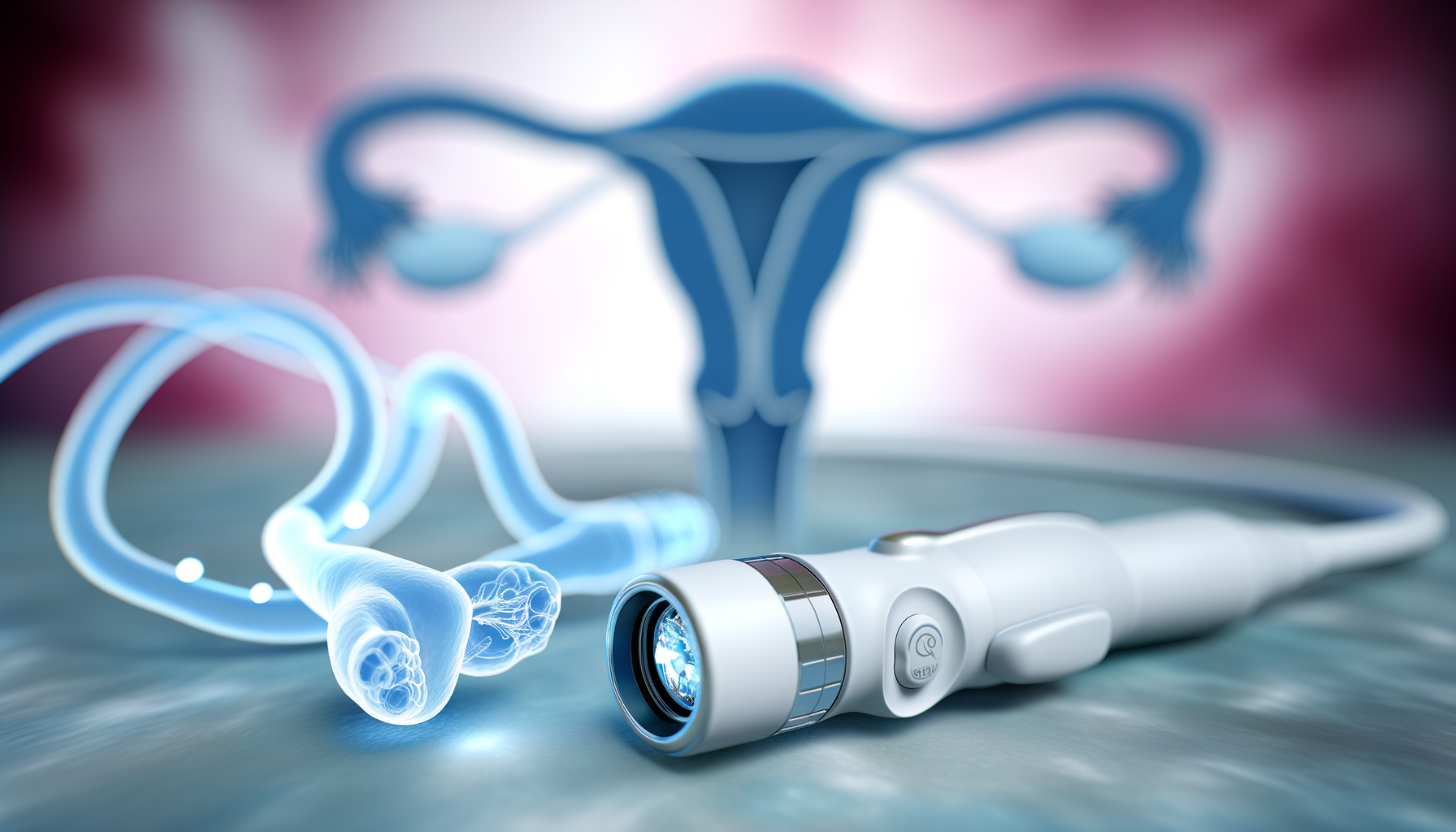
Ovarian cancer, known as the deadliest female reproductive malignancy, presents significant challenges in early detection due to its asymptomatic nature in initial stages. Dr. Jennifer Barton and her team at the University of Arizona have been at the forefront of developing a novel diagnostic tool, the falloposcope, to address this critical issue. The falloposcope is a minimally invasive endoscope designed to scan the fallopian tubes for early signs of ovarian cancer. The research behind the falloposcope is grounded in the understanding that the deadliest ovarian cancer phenotypes often originate in the fallopian tubes, where precursor lesions can remain undetected for years before metastasizing to the ovaries.
Design and Technological Innovations
The falloposcope represents a significant technological innovation, incorporating advanced imaging techniques such as optical coherence tomography and multispectral fluorescence imaging within a device just 0.8 millimeters in diameter. This “itty bitty” scope is capable of accurately detecting small lesions in the fallopian tubes without causing significant damage due to its small size. The design process involved multiple procedural and equipment refinements, resulting in a compact, robust, and quick imaging system optimized for patient imaging.
Pilot Human Trial and Initial Findings
Despite initial delays due to the COVID-19 pandemic, a pilot human trial was conducted with a cohort of 20 individuals undergoing fallopian tube removal for non-cancer reasons. The falloposcope successfully imaged sections of the fallopian tubes in most participants, demonstrating its capability to identify small lesions. These initial findings are promising, indicating that the falloposcope could play a crucial role in early intervention by detecting pre-invasive cells before they disseminate from the fallopian tubes to the ovaries.
FDA’s Stance and Safety Considerations
The U.S. Food and Drug Administration (FDA) has deemed the study to have “nonsignificant risk,” reflecting the safety profile of the falloposcope. The device’s testing on organs scheduled for removal inherently reduces risk further. However, the FDA’s designation does not diminish the importance of rigorous safety assessments and the need for continued refinement based on physician feedback and clinical trial outcomes. The successful and safe use of the falloposcope in the initial volunteers is a testament to the careful consideration of safety in its design and application.
Imaging Techniques Used by the Falloposcope
Fluorescence Imaging
Fluorescence imaging is a pivotal component of the falloposcope’s diagnostic capabilities. This technique leverages the properties of certain molecules within the tissue that absorb and emit light at different wavelengths. When illuminated with light of a specific wavelength, these molecules fluoresce, emitting light at a different wavelength. This emitted light is captured by the falloposcope, providing detailed information about the metabolic and functional state of the tissue. The ability to detect subtle changes in fluorescence can reveal the presence of precancerous or cancerous cells, which often exhibit altered metabolic processes compared to normal cells.
Optical Coherence Tomography
Optical Coherence Tomography (OCT) is another sophisticated imaging method employed by the falloposcope. OCT offers high-resolution, cross-sectional images of tissue structure by measuring the echo time delay and intensity of backscattered light. This non-invasive technique can visualize microscopic structural changes within the fallopian tubes that may indicate the early stages of cancer development. The falloposcope’s OCT system provides clinicians with a detailed view of the tissue architecture, enhancing the ability to distinguish between normal and abnormal tissue formations.
White Light Reflectance Imaging
White Light Reflectance Imaging (WLRI) is the third imaging modality integrated into the falloposcope. This technique involves shining white light onto the tissue and analyzing the reflected light spectrum. The characteristics of the reflected light can provide insights into the tissue’s composition and structure. WLRI is particularly useful for identifying surface-level abnormalities and can complement the deeper tissue analysis provided by OCT.
Comparative Analysis of Imaging Techniques
Each imaging technique utilized by the falloposcope offers unique advantages and contributes to a comprehensive assessment of fallopian tube health. Fluorescence imaging excels in detecting functional changes at the molecular level, while OCT provides a detailed view of tissue microstructure. WLRI adds to the diagnostic power by assessing surface-level tissue characteristics. Together, these modalities enable a multi-faceted evaluation of the fallopian tubes, significantly enhancing the early detection of ovarian cancer.
The integration of these imaging techniques into a single, minimally invasive device represents a significant advancement in gynecological diagnostics. By providing real-time, high-resolution images of the fallopian tubes, the falloposcope has the potential to revolutionize the early detection and treatment of ovarian cancer, ultimately improving patient outcomes and quality of life.
Implications of Early Detection on Treatment
Prophylactic Salpingo-Oophorectomies
Prophylactic salpingo-oophorectomies, the preventive removal of the ovaries and fallopian tubes, are a significant surgical intervention for women at high risk of developing ovarian cancer. This procedure is often recommended for those with genetic predispositions, such as BRCA1 or BRCA2 mutations. The rationale behind this surgery is the removal of potential sites where malignant transformations could occur, thereby reducing the risk of ovarian cancer development.
Potential Benefits and Drawbacks of Preventive Surgery
The benefits of prophylactic salpingo-oophorectomies are clear: a significant reduction in the risk of ovarian cancer and, consequently, a decrease in mortality associated with this disease. However, the drawbacks are not insignificant. Women undergoing this surgery face immediate menopause, with associated symptoms such as hot flashes and mood swings. Long-term consequences include increased risks of cardiovascular disease, osteoporosis, and potential negative impacts on sexual function and quality of life. Moreover, the psychological impact of such a drastic preventive measure cannot be overlooked.
Case Study Analysis of Preventive Measures
In a case study involving 122 patients with genetic predispositions to ovarian cancer, prophylactic salpingo-oophorectomies were performed. Post-surgical analysis of the removed tissues revealed that only a small fraction of these women were in the early stages of developing cancer. This finding suggests that while the surgery is effective in preventing cancer, it may also be an overtreatment for many, subjecting them to unnecessary surgical risks and the aforementioned long-term health consequences.
Alternative Approaches to Ovary Removal
Given the potential downsides of prophylactic surgery, alternative approaches are being explored. One such approach is regular screening with the falloposcope, which could allow for the monitoring of at-risk women without immediate surgery. This could potentially delay or even eliminate the need for ovary removal in some cases. Additionally, the development of medical therapies targeting early-stage lesions in the fallopian tubes could provide non-surgical options for cancer prevention. The goal is to balance the benefits of preventing ovarian cancer with the preservation of quality of life and overall health.
In conclusion, while prophylactic salpingo-oophorectomies offer a definitive preventive measure against ovarian cancer, the implications of early detection through technologies like the falloposcope could revolutionize treatment protocols. By providing a non-invasive means to monitor at-risk individuals, the falloposcope may reduce the need for surgery, thereby sparing women from the immediate and long-term consequences of ovary removal. As research progresses, the hope is to refine treatment options to offer personalized and less invasive strategies for ovarian cancer prevention.
By the way, something for you, a little gift!!!
I am just in the middle of publishing my book. It’s about How women can balance their hormones. One part is about food and diet, of course.
Follow this link and enter your email.
I will send you this part of the book for free once the book is published. It has many concrete, practical tips and recipes and will help you feel better during menopause or times of Big hormonal fluctuations.
Annette, Damiva Lead for Health & Wellness

Enhancing Quality of Life and Treatment Options
Regular Screening with the Falloposcope
The advent of the falloposcope introduces a transformative approach to ovarian cancer screening. By enabling regular, non-invasive screenings, this tiny device can monitor the health of the fallopian tubes over time. This is particularly beneficial for women at high risk of developing ovarian cancer, as it allows for close surveillance without the need for immediate, radical surgical interventions. The ability to detect early-stage cancer could mean more treatment options and a better prognosis, potentially increasing the five-year survival rate for ovarian cancer patients significantly.
Delaying Invasive Procedures
One of the most profound benefits of the falloposcope is its potential to delay invasive procedures such as prophylactic salpingo-oophorectomies. These procedures, while lifesaving, can induce premature menopause and lead to a host of side effects, including hot flashes, mood swings, and increased risk of heart and bone diseases. With the falloposcope, patients who might otherwise undergo early removal of their ovaries and fallopian tubes can opt to wait, preserving their hormonal balance and quality of life for a longer period.
Feedback Loop Between Surgeons and Engineers
The development of the falloposcope is a shining example of the synergy between medical and engineering disciplines. Surgeons like Heusinkveld provide invaluable feedback to the engineering team, ensuring that the device is not only effective in detecting abnormalities but also user-friendly in a clinical setting. This ongoing collaboration is crucial for refining the falloposcope, enhancing its functionality, and ultimately improving patient outcomes.
Patient Experiences and Hormonal Considerations
Understanding patient experiences is vital in the context of ovarian cancer risk reduction. Anecdotal evidence suggests that women who undergo ovary removal at a young age often report a decline in their overall well-being, even when on supplemental hormones. The falloposcope offers a chance to preserve natural hormonal function by delaying surgery until it is absolutely necessary. This approach could significantly improve the postoperative quality of life for women who would otherwise face the challenges of surgically induced menopause.
In conclusion, the falloposcope stands as a beacon of hope, not only for its potential to detect early-stage ovarian cancer but also for its role in enhancing the quality of life for at-risk women. By allowing for regular screenings, delaying invasive procedures, fostering a feedback loop between surgeons and engineers, and considering the hormonal well-being of patients, this groundbreaking device could redefine ovarian cancer treatment and prevention.
Establishing a Baseline for ‘Normal’ Fallopian Tubes
Goals for Imaging and Data Collection
The primary objective in the development of the falloposcope is not only to detect early-stage ovarian cancer but also to establish a comprehensive baseline of what constitutes ‘normal’ fallopian tube health. This baseline is crucial for differentiating between healthy and potentially cancerous tissues. The falloposcope’s high-resolution imaging capabilities allow for detailed visualization of the fallopian tubes, which are narrow ducts connecting the uterus to the ovaries. By collecting a wide range of images from volunteers undergoing salpingectomy for non-cancerous reasons, researchers aim to gather a diverse set of data that reflects the natural variation in fallopian tube anatomy and physiology.
Defining ‘Normal’ in Fallopian Tube Health
Defining ‘normal’ fallopian tube health is a complex task due to individual anatomical differences among women. The falloposcope provides a unique opportunity to observe the inner landscape of the fallopian tubes in unprecedented detail. By analyzing tissues from volunteers, researchers can identify common characteristics and patterns that signify a healthy state. This includes the appearance of the mucosal lining, the texture and color of the tissue, and the absence of any abnormal growths or irregularities.
Physician Feedback and Image Assessment
As the falloposcope is piloted, physician feedback is integral to refining the imaging process. Dr. John Heusinkveld, who has been instrumental in the device’s initial inpatient uses, provides critical insights into the usability and functionality of the falloposcope. This feedback loop between surgeons and engineers is essential for enhancing the device’s design and ensuring that the images captured are of the highest quality and clinical relevance. Physicians assess the images for clarity, resolution, and the ability to provide meaningful information about the health of the fallopian tubes.
Future Research Directions
Looking ahead, the research team envisions expanding the use of the falloposcope to a broader patient population, including those at high risk for ovarian cancer. The long-term goal is to integrate regular falloposcope screenings into standard gynecological care, potentially delaying or even preventing the need for prophylactic surgeries. As more data is collected, machine learning algorithms could be developed to assist in the interpretation of images, further refining the definition of ‘normal’ and improving early detection rates. The ongoing collaboration between clinicians and engineers will continue to play a pivotal role in the evolution of the falloposcope as a revolutionary tool in the fight against ovarian cancer.
Future Prospects and Impact on Ovarian Cancer Screening
Path to FDA Approval and Commercial Availability
The journey of the falloposcope from a research breakthrough to a widely available clinical tool is paved with regulatory and manufacturing milestones. The U.S. Food and Drug Administration (FDA) has already deemed the study involving the falloposcope as having “nonsignificant risk,” which is a promising start. However, comprehensive clinical trials are necessary to demonstrate the device’s efficacy and safety in a broader population. Once the device has proven its clinical value, the path to FDA approval will involve rigorous review processes. Post-approval, manufacturing scalability and distribution channels must be established to ensure commercial availability, making this innovative tool accessible to healthcare providers and patients.
Potential Changes to Screening Protocols
With the advent of the falloposcope, ovarian cancer screening protocols could undergo significant changes. Currently, there is no effective screening for early-stage ovarian cancer, leading to late diagnoses and poor survival rates. The falloposcope’s ability to image the fallopian tubes with high resolution could facilitate the development of new screening guidelines, particularly for women at high risk of developing ovarian cancer. Regular falloposcope screenings could become a part of routine gynecological care, allowing for earlier detection and intervention, potentially saving lives and reducing the need for invasive surgeries.
Long-Term Goals for Ovarian Cancer Prevention
The long-term goals for ovarian cancer prevention hinge on the ability to detect precancerous changes before they develop into full-blown cancer. By establishing what “normal” fallopian tubes look like and identifying early deviations from this baseline, the falloposcope could play a crucial role in preventive strategies. The device could also aid in monitoring high-risk patients, providing a less drastic alternative to prophylactic salpingo-oophorectomies, which carry significant side effects. Ultimately, the goal is to shift the paradigm from reactive treatment to proactive management of ovarian health.
Concluding Thoughts on the Falloposcope’s Role
The falloposcope represents a beacon of hope in the fight against ovarian cancer. As researchers continue to refine the device and gather more data, its potential to revolutionize early detection and prevention is becoming increasingly clear. The falloposcope’s role could extend beyond diagnostics, influencing treatment decisions, patient quality of life, and long-term health outcomes. While the path to widespread clinical use is complex and requires further validation, the promise of the falloposcope in enhancing ovarian cancer screening is undeniable. As we look to the future, the falloposcope stands as a testament to the power of innovation in transforming patient care and advancing women’s health.


